Sickle Cell Disease Researchers Identify a Children’s Stroke Prediction Tool.

BY PATRICIA INACIO, PHD
Sickle Cell Disease Researchers Identify a Children’s Stroke Prediction Tool
The amount of oxygen the blood loses when traveling through the brain could help identify children with sickle cell disease who are at risk of having a stroke, a study reports.
Scientists call the measurement the oxygen extraction fraction because it refers to the percentage of oxygen that the brain removes from blood.
The study, “Regional oxygen extraction predicts border zone vulnerability to stroke in sickle cell disease,” was published in the journal Neurology.
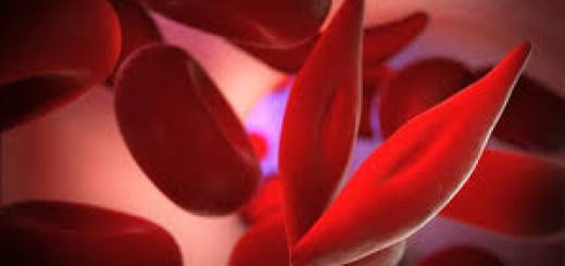
A hallmark of sickle cell disease is an accumulation of sickle-shaped red blood cells instead of the normal oval-shaped ones. The abnormal cells increase the risk that blood will flow slower through several organs, including bones, lungs, spleen, central nervous system, the eye’s retina, and the kidneys.
Researchers wanted to know if the risk of a brain infarction — or obstruction of blood supply to the brain — was higher in children with sickle cell disease than in healthy children. The common name for a brain infarction is a stroke.
The study involved 56 children — 36 with sickle cell disease and 20 controls. Researchers used MRI scans to obtain three blood-flow measurements in the children’s brains. The three were voxel-wise cerebral blood flow, oxygen extraction fraction, and cerebral metabolic rate of oxygen utilization.
Oxygen extraction fraction is an important measure because the higher it is, the greater the risk of a blood vessel in the brain being blocked.
Researchers discovered that children with sickle cell disease had poorer scores on all three blood-flow measures. The oxygen extraction fraction was 50 percent higher in children with the disease than in controls. It was 43 percent in the patients and 29 percent in the healthy children.
The research team was able to pinpoint an area of the sickle cell disease patients’ brains where the oxygen extraction fraction was highest — a deep white matter region. It correlated with areas of increased risk of stroke found in another study of children with sickle cell disease.
Overall, the findings showed that “elevated OEF [oxygen extraction fraction] in the deep white matter identifies a signature of metabolically stressed brain tissue at increased stroke risk in pediatric patients” with sickle cell disease, the researchers wrote.
The team plans to continue following the sickle cell disease patients to see whether increased levels of oxygen extraction fraction in various regions of their brains can help predict their risk of stroke.
Post Credit :
Patricia Inacio, PhD
Patricia holds her Ph.D. in Cell Biology from University Nova de Lisboa, and has served as an author on several research projects and fellowships, as well as major grant applications for European Agencies. She also served as a PhD student research assistant in the Laboratory of Doctor David A. Fidock, Department of Microbiology & Immunology, Columbia University, New York.


Recent Comments